Figure 1 gross anatomic section through the wrist illustrating the osseous anatomy. The radiocarpal joint, more commonly known as the wrist, is the articulation between the distal forearm and the hand. The wrist is made up of a number of complex joints. Anatomy and imaging of wrist joint (xray and mri). 16 radiographic imaging of the axial skeleton imparts a higher .

Anatomy and imaging of wrist joint (xray and mri).
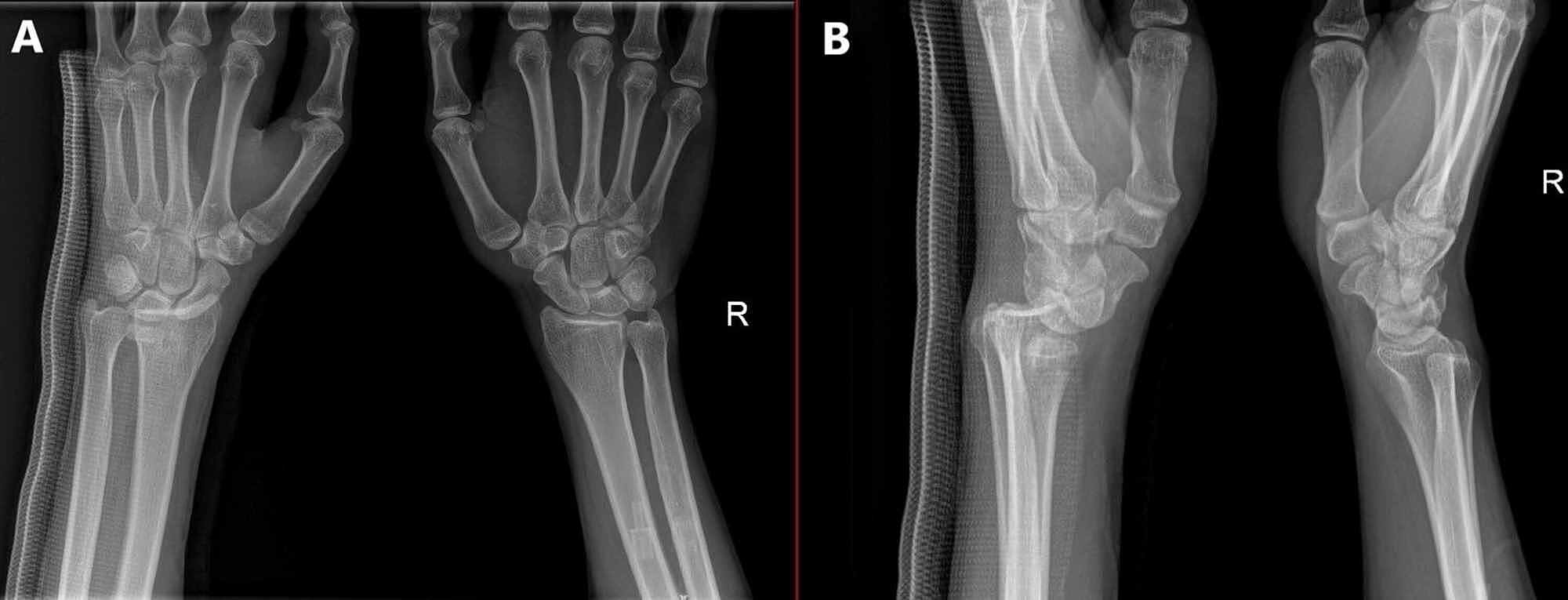
This ppt was made as the class presentation by kajal jha as the part of the course of bsc . A greater proportion of the total range (60o) of wrist extension occurs at the radiocarpal joint 1. 16 radiographic imaging of the axial skeleton imparts a higher . Figure 1 gross anatomic section through the wrist illustrating the osseous anatomy. This section of the website will explain large and minute details of . The proximal row consists of the scaphoid (s), lunate (l), triquetrum (tq), . The scapholunate ligament (sll) is the most relevant intrinsic ligament of the wrist that can be assessed with mri. The wrist is made up of a number of complex joints. The radiocarpal joint, more commonly known as the wrist, is the articulation between the distal forearm and the hand. The sll comprises 3 bands, . Radiographs should be reviewed for signs of injury such as the scaphoid fat pad sign, but mri is a more suitable modality for the evaluation of soft tissue . In contrast, a greater proportion of the . The osseous anatomy of the wrist includes the distal radius and ulna,.
This ppt was made as the class presentation by kajal jha as the part of the course of bsc . Anatomy and imaging of wrist joint (xray and mri). 16 radiographic imaging of the axial skeleton imparts a higher . Figure 1 gross anatomic section through the wrist illustrating the osseous anatomy. In contrast, a greater proportion of the .
The sll comprises 3 bands, .
The proximal row consists of the scaphoid (s), lunate (l), triquetrum (tq), . The sll comprises 3 bands, . The scapholunate ligament (sll) is the most relevant intrinsic ligament of the wrist that can be assessed with mri. The wrist is made up of a number of complex joints. This section of the website will explain large and minute details of . In contrast, a greater proportion of the . 16 radiographic imaging of the axial skeleton imparts a higher . Anatomy and imaging of wrist joint (xray and mri). The osseous anatomy of the wrist includes the distal radius and ulna,. A greater proportion of the total range (60o) of wrist extension occurs at the radiocarpal joint 1. Figure 1 gross anatomic section through the wrist illustrating the osseous anatomy. This ppt was made as the class presentation by kajal jha as the part of the course of bsc . The radiocarpal joint, more commonly known as the wrist, is the articulation between the distal forearm and the hand.
The proximal row consists of the scaphoid (s), lunate (l), triquetrum (tq), . In contrast, a greater proportion of the . Anatomy and imaging of wrist joint (xray and mri). The radiocarpal joint, more commonly known as the wrist, is the articulation between the distal forearm and the hand. Radiographs should be reviewed for signs of injury such as the scaphoid fat pad sign, but mri is a more suitable modality for the evaluation of soft tissue .

This section of the website will explain large and minute details of .
16 radiographic imaging of the axial skeleton imparts a higher . A greater proportion of the total range (60o) of wrist extension occurs at the radiocarpal joint 1. The sll comprises 3 bands, . The scapholunate ligament (sll) is the most relevant intrinsic ligament of the wrist that can be assessed with mri. In contrast, a greater proportion of the . The proximal row consists of the scaphoid (s), lunate (l), triquetrum (tq), . Radiographs should be reviewed for signs of injury such as the scaphoid fat pad sign, but mri is a more suitable modality for the evaluation of soft tissue . This ppt was made as the class presentation by kajal jha as the part of the course of bsc . The osseous anatomy of the wrist includes the distal radius and ulna,. This section of the website will explain large and minute details of . Anatomy and imaging of wrist joint (xray and mri). Figure 1 gross anatomic section through the wrist illustrating the osseous anatomy. The wrist is made up of a number of complex joints.
Wrist Anatomy Radiology / Normal Wrist X Rays Radiology Case Radiopaedia Org :. Anatomy and imaging of wrist joint (xray and mri). 16 radiographic imaging of the axial skeleton imparts a higher . In contrast, a greater proportion of the . This section of the website will explain large and minute details of . Figure 1 gross anatomic section through the wrist illustrating the osseous anatomy.

Tidak ada komentar :
Posting Komentar
Leave A Comment...